Pelger-Huët anomaly is a hereditary disorder of the leukocytes characterized by granulocytes with hyposegmented nuclei and a coarse, mature pattern of chromatin. The morphology of the granulocyte nuclei may be round, oval, dumbbell-shaped, peanut-shaped, or bilobulate, or they may appear as a band. Pelger-Huët anomaly has been reported in human beings, rabbits, cats, dogs, horses and mice. In dogs, the condition has been described in a number of breeds, including the Cocker Spaniel, Basenji, Border Collie, English and American Foxhounds, Samoyed, Australian Shepherd, Australian Cattle Dog, Boston Terrier, German Shepherd Dog, coonhound, and mixed-breed dogs. Pelger-Huët anomaly is usually a rare disease but is more common in some breeds such as Australian Shepherds where an estimated incidence of 9.8% has been reported.
Pelger-Huët anomaly is an autosomal dominant hereditary anomaly, except in Australian Shepherds where incomplete penetrance has been reported. In human beings, the anomaly has been attributed to a mutation in the lamin B receptor gene. This receptor is an inner nuclear membrane protein responsible for the traffic of heterochromatin and lamins to the nuclear Membrane. Its disruption causes abnormalities in nuclear heterochromatin and, consequently, in nuclear morphology as described previously.
It is generally accepted that immunodeficiency and predisposition to infection do not occur in patients with Pelger-Huët anomaly. The comparison of the function of Pelger-Huët neutrophils with normal neutrophils of dogs revealed no significant differences in neutrophil adherence, random movement, chemotaxis, phagocytosis, and bactericidal activity. Other studies also demonstrated that Pelger-Huët leukocytes function similarly to normal cells.
The practical significance of recognizing Pelger-Huët anomaly is to distinguish it from severe infection and to thereby avoid unnecessary treatment, further diagnostic testing, and associated medical expenses. This differentiation can be made through blood smear evaluation by observation of the appearance of the nuclear lobes, nuclear size, and chromatin pattern. Demonstrating similar nuclear hyposegmentation in relatives such as the sire, dam, and/or siblings is crucial for a definitive diagnosis of Pelger-Huët anomaly
Literature:
Pelger-Huët anomaly in two related mixed-breed dogs, DOI: 10.1177/1040638711407891
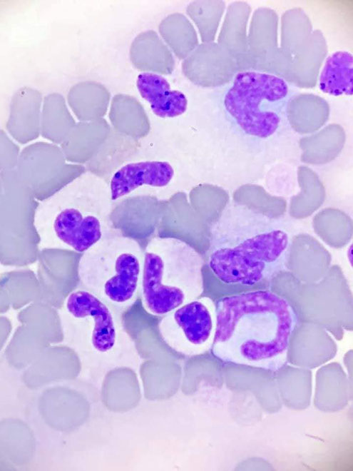
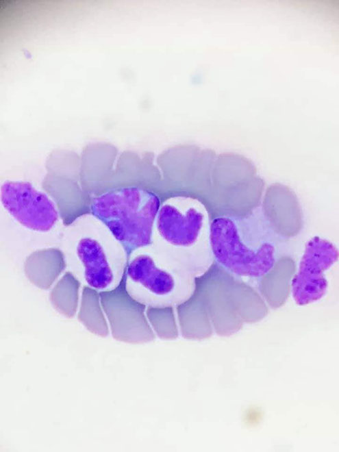
Blood smear, mod. Romanowsky stain, 100x oil
Description:
- neutrophil granulocytes with hyposegmented nuclei (bands, metamyelocytes)
- coarse, mature pattern of chromatin
- no signs of toxicity (inflammation)